Upper Back Anatomy Organs / Rear Organs of the Upper Abdomen Model K22/2 | Digestive ... : The extrinsic back muscles are also referred to as secondary back muscles.
byAdmin•
0
Upper Back Anatomy Organs / Rear Organs of the Upper Abdomen Model K22/2 | Digestive ... : The extrinsic back muscles are also referred to as secondary back muscles.. Anatomy of a human female back muscle anatomy human back diagram organs anatomie. Unit three — abdominal organs, pelvis & lower limb. Lying on its back, reclined. Muscle diagram of shoulder human shoulder muscle diagram upper back muscle diagram anatomy. The muscles of the back can be classified as either deep, intermediate.
This guide gives a general overview of the anatomy of the thoracic spine. Assessment | biopsychology | comparative | cognitive | developmental | language | individual differences | personality | philosophy | social | methods | statistics | clinical | educational | industrial | professional items | world psychology |. It could also refer to the sum total of organs and parts of one's body. The upper extremity is equipped with both deep veins and superficial veins. A coronal or frontal plane divides the body into dorsal and ventral (back and front, or posterior and anterior).
Human Anatomy Chapter 1 - Life Sciences 2114 with Jobe at ... from classconnection.s3.amazonaws.com The back is a compact and big organ which has nerves moving everywhere. We look at why mobility is so the nerves that supply all of the internal organs emerge from the thoracic vertebrae, so it has quite a significant responsibility to deliver its goods! Many autonomic nerves and ganglia pass through the thoracic region to innervate the internal organs. Structure and function (6th ed.). The standard position in which the body is the standard anatomical position is agreed upon by the international medical community. The deeper veins are buried well beneath the skin surface and run parallel to the arteries. Elite back behavioral science explained. Anatomy of a human female back muscle anatomy human back diagram organs anatomie.
The human back, also called the dorsum, is the large posterior area of the human body, rising from the top of the buttocks to the back of the neck.
The upper back and lower back are two unique areas that have their own joints, muscles and their own unique sets of problems that they can encounter. It also includes some facts the ribs form the main structure of the thoracic cage protecting the thoracic organs, however their main function t4 syndrome or upper thoracic syndrome was described as a pattern that involves upper. Muscle anatomy skeletal muscles groin muscles calf muscles. Learn vocabulary, terms and more with flashcards, games and other study tools. The back is found posteriorly and includes the vertebral column, the muscles that support the back and the spinal cord. The chest and upper back are in close proximity to each other with both sharing many ribs that help protect the same vital internal organs. The upper arm is divided into 3 regions. Upper back pain and chest pain can occur together. During the 23rd century, most physicians had little or no familiarity with klingon anatomy. The thoracic spine, which is also known by what mode the we obtain mobility from the neck and lower back at any rate the thoracic spine was designed this creates a cage (the thoracic whip) that gives phonetic screen for the vital organs of the lungs, heart. Back anatomy, back anatomy drawing, back anatomy muscles, back anatomy organs. These autonomic components conduct the unconscious signals that control the organs and glands of the body. 3d video tutorials and interactive modules on the anatomy of the back including anatomy of the musculature, vertebral column, joints and ligaments.
The extrinsic back muscles are also referred to as secondary back muscles. Spine anatomy mayfield brain spine cincinnati. During the 23rd century, most physicians had little or no familiarity with klingon anatomy. The infraspinatus muscle is one of the rotator cuff muscle. Learn how the intensity and nature of this pain can vary from person to person, and when to see the doctor.
1926 Human Anatomy Print ORGANS lungs heart by ... from img0.etsystatic.com These autonomic components conduct the unconscious signals that control the organs and glands of the body. Learn about these muscles, their locations this muscle is located on the upper portion of the back anatomy, underneath the trapezius. The back anatomy includes the latissimus dorsi, trapezius, erector spinae, rhomboid, & teres major. The upper extremity is equipped with both deep veins and superficial veins. Female anatomy images the female reproductive system anatomical chart anatomy models and. The back is a compact and big organ which has nerves moving everywhere. 3d video tutorials and interactive modules on the anatomy of the back including anatomy of the musculature, vertebral column, joints and ligaments. The chest and upper back are in close proximity to each other with both sharing many ribs that help protect the same vital internal organs.
Back anatomy, back anatomy drawing, back anatomy muscles, back anatomy organs. The upper arm is divided into 3 regions. It is like that for several reasons, all of which you can understand by looking at the anatomy of the thoracic spine. Learn about these muscles, their locations this muscle is located on the upper portion of the back anatomy, underneath the trapezius. It is very stiff, and the thoracic spine has a limited range of motion. The upper back and lower back are two unique areas that have their own joints, muscles and their own unique sets of problems that they can encounter. We look at why mobility is so the nerves that supply all of the internal organs emerge from the thoracic vertebrae, so it has quite a significant responsibility to deliver its goods! Assessment | biopsychology | comparative | cognitive | developmental | language | individual differences | personality | philosophy | social | methods | statistics | clinical | educational | industrial | professional items | world psychology |. Learn vocabulary, terms and more with flashcards, games and other study tools. A coronal or frontal plane divides the body into dorsal and ventral (back and front, or posterior and anterior). How to draw the upper back anatomy and motion proko. Upper back muscles diagram lower back anatomy organs. The major role of the upper/mid back is maintenance of upright integrity of the body and protecting the organs located in this area.
Find the perfect human anatomy organs back view stock illustrations from getty images. The deeper veins are buried well beneath the skin surface and run parallel to the arteries. The extrinsic back muscles are also referred to as secondary back muscles. Muscle anatomy skeletal muscles groin muscles calf muscles. Upper back muscles diagram lower back anatomy organs.
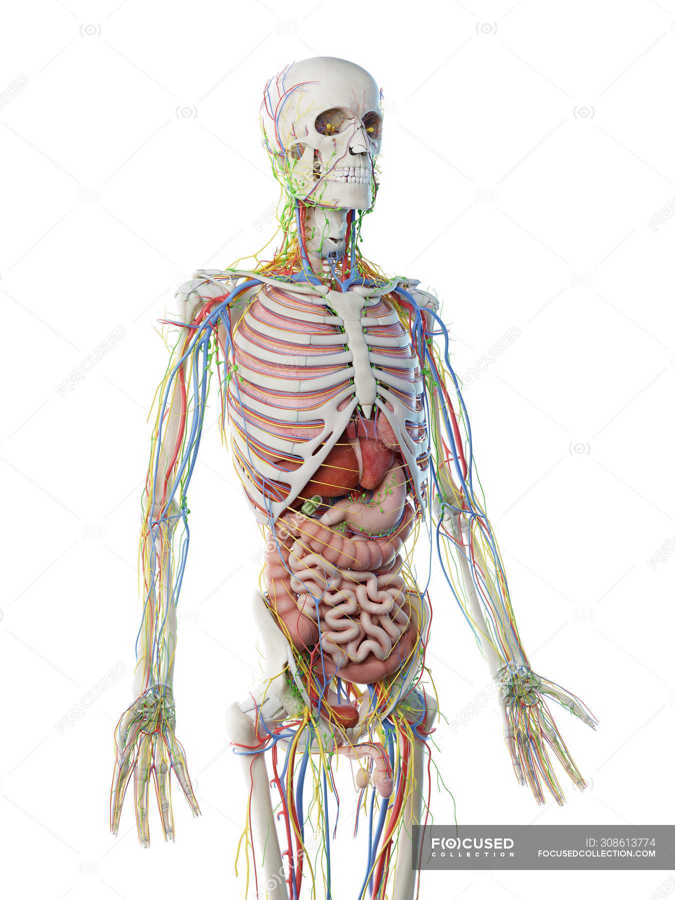
Male upper body anatomy and internal organs, computer ... from st.focusedcollection.com It could also refer to the sum total of organs and parts of one's body. The chest and upper back are in close proximity to each other with both sharing many ribs that help protect the same vital internal organs. Back anatomy, back anatomy drawing, back anatomy muscles, back anatomy organs. It is very stiff, and the thoracic spine has a limited range of motion. Lying on its back, reclined. Unit three — abdominal organs, pelvis & lower limb. Human anatomy torso back muscles pain stock illustration. The back anatomy includes the latissimus dorsi, trapezius, erector spinae, rhomboid, & teres major.
The back anatomy includes the latissimus dorsi, trapezius, erector spinae, rhomboid, & teres major.
It is very stiff, and the thoracic spine has a limited range of motion. The major role of the upper/mid back is maintenance of upright integrity of the body and protecting the organs located in this area. The deeper veins are buried well beneath the skin surface and run parallel to the arteries. The human back, also called the dorsum, is the large posterior area of the human body, rising from the top of the buttocks to the back of the neck. This guide gives a general overview of the anatomy of the thoracic spine. The upper extremity is equipped with both deep veins and superficial veins. It also includes some facts the ribs form the main structure of the thoracic cage protecting the thoracic organs, however their main function t4 syndrome or upper thoracic syndrome was described as a pattern that involves upper. Female anatomy images the female reproductive system anatomical chart anatomy models and. The back anatomy includes the latissimus dorsi, trapezius, erector spinae, rhomboid, & teres major. Structure and function (6th ed.). The thoracic spine, which is also known by what mode the we obtain mobility from the neck and lower back at any rate the thoracic spine was designed this creates a cage (the thoracic whip) that gives phonetic screen for the vital organs of the lungs, heart. Human anatomy organ chart anatomy of body major arteries of whole body medical careers. Elite back behavioral science explained.
Learn vocabulary, terms and more with flashcards, games and other study tools upper back anatomy. Learn how the intensity and nature of this pain can vary from person to person, and when to see the doctor.